
Focused ultrasound represents a different way of treating prostate tissue. Rather than using sound waves only to create diagnostic images, therapeutic ultrasound concentrates acoustic energy within a carefully selected area. The resulting heat can ablate targeted tissue while limiting energy exposure to structures outside the treatment zone. At Texas Prostate in Farmers Branch, TX, within the Dallas-Fort Worth Metroplex, focused ultrasound technology provides the context for understanding modern approaches to treating localized prostate cancer and benign prostatic hyperplasia, or BPH.
Although focused ultrasound systems may use different delivery methods and forms of image guidance, they share several fundamental principles. A treatment plan identifies the tissue to be addressed, ultrasound energy is directed toward that region, and temperature or imaging feedback helps the physician monitor the process. Understanding these principles can make an unfamiliar technology easier to visualize.
From Diagnostic Imaging to Therapeutic Energy
Most people associate ultrasound with imaging. During a diagnostic ultrasound examination, a transducer sends sound waves into the body and receives returning echoes. A computer interprets those echoes to create an image of internal anatomy. The sound energy used for diagnostic imaging is relatively low and is not intended to heat or destroy tissue.
Therapeutic ultrasound uses the same broad concept of sound-wave transmission but operates at energy levels designed to produce a biological effect. The waves are delivered so that their energy converges on or is directed toward a planned treatment area. This concentration of energy creates heat within the selected tissue. Once the tissue reaches an adequate temperature for a sufficient amount of time, its cells undergo thermal coagulation and can no longer function normally.
The process is often compared to using a magnifying glass to concentrate sunlight. Light passing through the glass is not equally intense everywhere; it becomes strongest at the focal point. With focused ultrasound, acoustic energy is similarly controlled so that the most significant heating occurs within the intended treatment zone rather than uniformly along the entire path of the sound waves.
How Ultrasound Energy Ablates Prostate Tissue
Ablation is the controlled destruction of tissue. In prostate treatment, focused or directional ultrasound creates heat that produces coagulative necrosis within a prescribed region. Proteins inside the targeted cells denature, cell membranes lose their integrity, and the treated tissue gradually breaks down as part of the body’s natural healing response.
Temperature is only one part of the equation. The duration of heating also matters. A very high temperature may create the intended tissue effect within seconds, while a lower temperature may require a longer exposure. Treatment systems therefore regulate factors such as ultrasound power, frequency, applicator movement, and exposure time to deliver an appropriate thermal dose.
The prostate is located near the urethra, rectum, urinary sphincter, bladder neck, and nerves associated with sexual function. This compact anatomy makes planning especially important. The purpose of image-guided treatment is not simply to generate heat. It is to shape and control the heated region so that it corresponds as closely as possible to the physician-defined treatment boundary.
The Importance of Treatment Planning and Image Guidance
Focused ultrasound begins with detailed information about the prostate and the area being considered for treatment. Magnetic resonance imaging, ultrasound imaging, biopsy results, prostate-specific antigen measurements, prostate size, and the location of abnormal tissue may all contribute to clinical planning. The exact evaluation depends on the condition being addressed and the technology being considered.
During treatment, image guidance helps establish the location of the prostate, applicator, target, and nearby anatomy. It also helps the physician confirm that energy is being directed into the planned region. Some systems use ultrasound images to guide treatment, while others incorporate magnetic resonance imaging and real-time temperature mapping.
Imaging is especially important because the physician cannot directly see the tissue response in the same way that exposed tissue could be viewed during an open procedure. Instead, the system supplies information about anatomy, applicator position, energy delivery, and temperature changes. This creates a feedback-driven process in which treatment can be planned, observed, and adjusted.
Two Routes for Delivering Ultrasound Energy
The prostate can be reached with therapeutic ultrasound through different anatomical routes. In HIFU, or high-intensity focused ultrasound, energy is commonly delivered through a transrectal probe. The probe sends multiple ultrasound waves toward a focal point inside the prostate. At that point, their combined intensity creates enough heat to ablate the selected tissue.
The physician treats a planned area by moving the focal point through a sequence of adjacent locations. Each application produces a small ablation zone. Together, these overlapping zones form the larger treatment volume. Imaging helps the physician maintain alignment between the planned target and the location where energy is being deposited.
The TULSA Procedure uses a different route. TULSA stands for Transurethral Ultrasound Ablation. An ultrasound applicator is positioned within the urethra, placing the energy source inside the prostate rather than outside it. Directional ultrasound is transmitted outward from the applicator toward the prostate tissue selected for ablation.
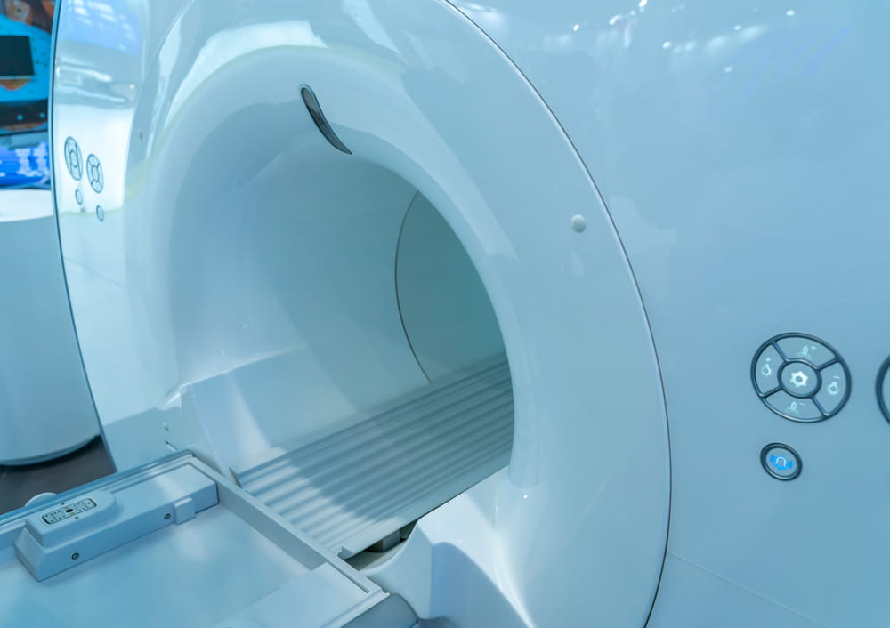
TULSA is performed with magnetic resonance imaging guidance and MRI thermometry. The applicator rotates as it delivers energy, while software uses temperature information to regulate factors such as power and rotation. Cooling within the applicator helps protect the urethra, and a separate cooling device helps limit heat exposure to the rectum.
How Real-Time Temperature Feedback Improves Control
A major challenge in thermal ablation is that living tissue does not heat identically in every person. Blood flow can carry heat away from an area, tissue composition can influence energy absorption, and prostate calcifications may reflect or interfere with ultrasound transmission. Treatment systems must account for these variations rather than relying only on a predetermined energy setting.
MRI thermometry estimates temperature changes throughout the treatment region. Instead of merely showing where the prostate is located, it provides ongoing feedback about how the tissue is responding to energy. Closed-loop software can compare the measured temperature pattern with the physician’s treatment plan and automatically modify energy delivery.
This feedback helps prevent two undesirable outcomes: insufficient heating within the target and unintended heating beyond it. If a region is warming too slowly, treatment parameters may be adjusted to deliver a sufficient thermal dose. If heat approaches a protected boundary, the system may reduce or redirect energy. The objective is a controlled treatment volume rather than the indiscriminate heating of the entire surrounding area.
Focal, Partial-Gland, and Whole-Gland Treatment Plans
Focused ultrasound technology can be configured for different treatment patterns. Focal ablation addresses a defined lesion or limited region. Partial-gland treatment covers a larger portion of the prostate, such as one side or another physician-defined volume. Whole-gland ablation treats nearly all planned prostate tissue while preserving specified safety margins around sensitive anatomy.
The appropriate treatment volume is not determined by technology alone. It depends on the diagnosis, the number and location of clinically significant lesions, prostate size, previous treatments, urinary symptoms, imaging findings, and individual priorities. A highly precise energy source does not eliminate the need for careful patient selection or diagnostic evaluation.
Precision also does not mean that ultrasound energy affects only abnormal cells. The technology cannot independently distinguish a cancer cell from a healthy prostate cell at the microscopic level. Instead, the physician defines a treatment boundary based on available diagnostic information, and the system ablates the tissue located within that boundary.
What Happens After Thermal Ablation
Immediately after focused ultrasound treatment, the prostate may develop temporary swelling and inflammation. Because the urethra passes through the gland, this response can affect urinary flow. A urinary catheter may therefore be used for a period while the prostate begins to recover.
The ablated tissue does not disappear instantly. Over time, the body removes damaged cells and reorganizes the treated area. Urinary symptoms, changes in urinary frequency, blood in the urine, pelvic discomfort, or temporary irritation may occur during recovery. The nature and duration of these effects can vary according to the amount and location of tissue treated.
Follow-up remains important after the initial recovery period. Depending on the original diagnosis, monitoring may include prostate-specific antigen testing, imaging, symptom evaluation, urinary testing, or a follow-up biopsy. The broader patient experience can include preparation, the treatment day, catheter management, recovery instructions, and ongoing surveillance rather than a single isolated procedure.
Understanding the Technology in Context
Focused ultrasound combines several technologies that work together: an energy-producing applicator, detailed imaging, computerized treatment planning, temperature or positioning feedback, and protective cooling systems when applicable. Its precision comes from the coordination of these components, not from sound energy alone.
It is also important to distinguish technical capability from individual suitability. Prostate anatomy, calcifications, gland size, disease location, cancer risk category, urinary function, previous treatment, and overall health can all influence whether a particular ultrasound approach is appropriate. A complete evaluation is needed to interpret those factors in relation to the goals and limitations of treatment.
Focused ultrasound is best understood as image-guided thermal ablation: sound energy is delivered through a controlled pathway, concentrated within a physician-defined region, and monitored as it creates a therapeutic temperature. For people researching prostate cancer or BPH care in the Dallas-Fort Worth Metroplex, Texas Prostate in Farmers Branch, TX, provides a local point of reference for learning how this technology is applied to prostate tissue.
Resources
Chin, J. L., Billia, M., Relle, J., Roethke, M. C., Popeneciu, I. V., Kuru, T. H., & Schlemmer, H. P. (2024). MR-Guided Transurethral Ultrasound Ablation (TULSA)—An Emerging Focal Therapy for Localized Prostate Cancer. Cardiovascular and Interventional Radiology.
Dababou, S., Marrocchio, C., Scipione, R., Erasmus, H. P., Ghanouni, P., Anzidei, M., Catalano, C., & Napoli, A. (2018). High-Intensity Focused Ultrasound for the Treatment of Prostate Cancer: A Review. Journal of Personalized Medicine.
U.S. Food and Drug Administration. (2023). TULSA-PRO System: 510(k) Premarket Notification K230692. U.S. Department of Health and Human Services.