Men researching focused ultrasound treatment for prostate conditions may encounter two frequently discussed technologies: TULSA Pro® and HIFU. Both use concentrated ultrasound energy to heat selected prostate tissue, but they differ significantly in how the energy reaches the prostate, how treatment is planned, and how temperature is monitored during the procedure. For patients in the Dallas-Fort Worth Metroplex, Texas Prostate in Farmers Branch, TX, provides a local point of reference for understanding these emerging prostate treatment technologies.
The Shared Principle Behind Focused Ultrasound
Focused ultrasound treatments use sound-wave energy rather than an incision to create controlled heat within targeted tissue. When the ultrasound energy is concentrated at a specific location, the temperature rises enough to cause thermal ablation. The objective is to treat the planned area while limiting unnecessary exposure to nearby structures.
This general principle is used by both TULSA Pro® and HIFU. However, the fact that both methods use ultrasound does not make them interchangeable. Their delivery routes, imaging environments, treatment patterns, and monitoring capabilities create distinctly different procedural experiences.
The suitability of either approach depends on more than the presence of a prostate condition. Prostate size, disease location, imaging findings, biopsy results, urinary function, previous treatment, anatomy, and overall health may all influence whether focused ultrasound is appropriate and which technique deserves consideration.
How TULSA Pro® Delivers Ultrasound Energy
TULSA stands for Transurethral Ultrasound Ablation. During this procedure, a slender ultrasound applicator is positioned within the urethra, which passes through the center of the prostate. The device directs ultrasound energy outward from inside the prostate toward the planned treatment boundary.
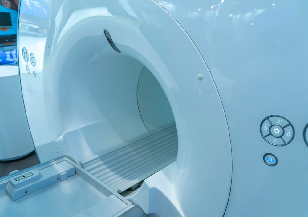
TULSA Pro is performed within an MRI environment. Magnetic resonance imaging provides detailed anatomical visualization, assists with treatment planning, and allows the treatment team to observe temperature changes as therapy progresses. The system uses real-time MRI thermometry to measure heat within and around the targeted tissue.
Because the applicator delivers energy from the inside outward, treatment can be customized around the prostate’s shape and the location of the intended treatment area. Depending on the clinical situation, a treatment plan may involve a smaller targeted region, a larger portion of the gland, or nearly the entire prostate while deliberately protecting selected structures.
Cooling is also an important component of the procedure. The urethra and rectum are actively cooled to help protect these structures from unintended heat. The treatment system continuously adjusts energy delivery based on temperature feedback and the boundaries established during planning.
How Transrectal HIFU Reaches the Prostate
HIFU stands for high-intensity focused ultrasound. In transrectal HIFU, an ultrasound probe is positioned in the rectum next to the prostate. The device directs multiple ultrasound beams through the rectal wall and focuses them at selected points inside the gland.
At the focal point, the concentrated energy produces a rapid increase in temperature that destroys the intended tissue. The probe is repositioned systematically so that multiple adjacent treatment points can be created throughout the planned area. This allows HIFU to treat an individual lesion, a portion of the prostate, or a more extensive area when clinically appropriate.
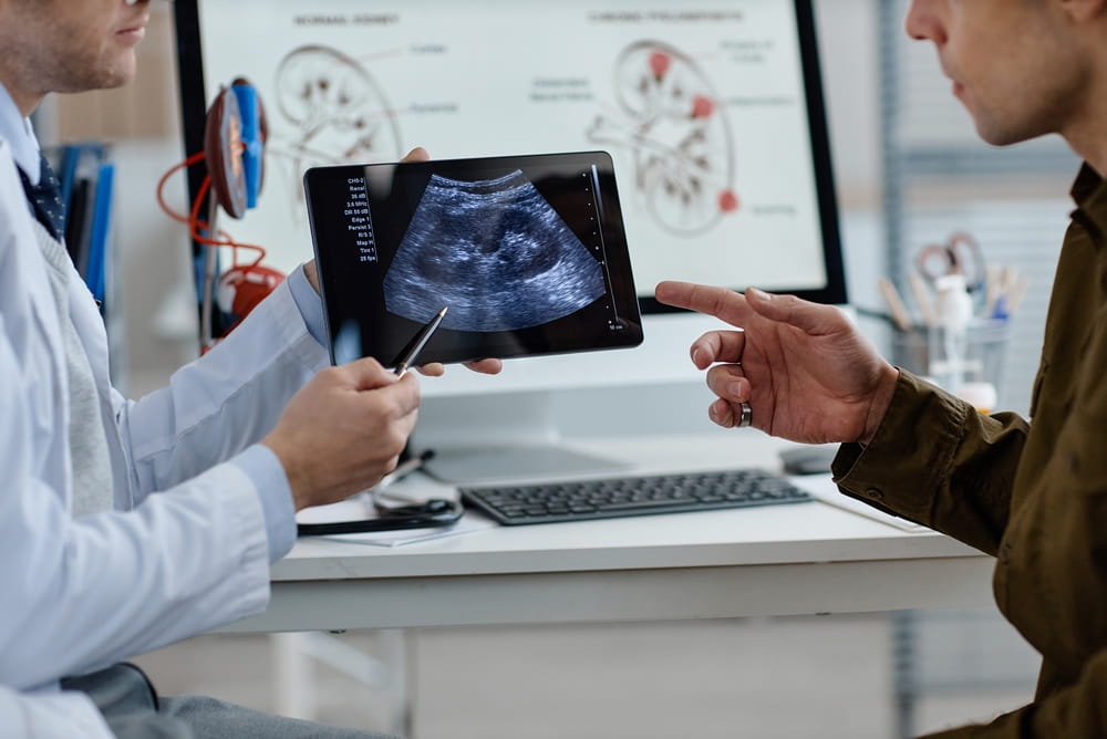
Transrectal HIFU commonly uses ultrasound imaging to visualize the prostate and guide treatment. Some systems can also incorporate information from magnetic resonance imaging, biopsy mapping, or image-fusion technology during the planning process. Unlike TULSA Pro, however, conventional transrectal HIFU is not performed with continuous MRI-based temperature mapping throughout the procedure.
The rectal delivery route also affects which parts of the prostate may be easier or more difficult to reach. Prostate dimensions, calcifications, treatment depth, rectal anatomy, and the location of suspicious tissue can influence procedural planning.
MRI Thermometry Is a Major Difference
One of the clearest distinctions between the two technologies is how heat is monitored. TULSA Pro uses MRI thermometry to create continually updated temperature maps during treatment. These maps show where tissue has reached the planned therapeutic temperature and where additional energy may be needed.
The system can respond to this feedback by modifying ultrasound power and adjusting the rotation or positioning of the applicator. This closed-loop approach is intended to keep the thermal boundary aligned with the physician’s treatment plan, even when tissue characteristics cause heat to spread differently than initially predicted.
HIFU treatment is guided primarily through ultrasound visualization and established energy-delivery patterns. The physician plans a series of overlapping focal points and monitors the prostate with the treatment system’s imaging capabilities. Although planning and monitoring technologies continue to evolve, traditional transrectal HIFU does not provide the same continuous MRI temperature feedback used during TULSA.
This difference does not establish that one technology is automatically preferable in every case. It identifies a meaningful distinction in how the treatment team observes and controls the ablation process.
Treatment Direction and Access Shape the Procedure
The direction in which ultrasound travels also separates the two treatments. TULSA Pro sends directional energy outward from the urethra. Transrectal HIFU sends focused energy forward from the rectal probe into the prostate.
An inside-out approach may provide flexibility when shaping treatment around the gland’s contours. An outside-in approach can offer direct transrectal access to selected prostate regions. The relevance of these differences depends on the location of the tissue being treated and its relationship to the urethra, rectum, urinary sphincter, neurovascular bundles, and prostate capsule.
Prostate calcifications may be particularly important because calcium can interfere with the transmission of ultrasound energy. Their size and location may affect treatment planning differently depending on whether the sound waves originate in the urethra or rectum.
Neither procedure can be selected responsibly from a single MRI image or PSA result. A complete assessment generally considers multiple diagnostic findings and the technical ability to reach the intended tissue without creating unacceptable risk to adjacent structures.
Focal and Whole-Gland Treatment Strategies
Both technologies may be discussed in connection with focal, partial-gland, or whole-gland treatment. These terms describe how much prostate tissue is included in the planned ablation rather than identifying a specific device.
Focal treatment concentrates on a defined area of clinically significant disease while preserving more untreated prostate tissue. Partial-gland treatment may include a larger region, such as one side of the prostate. Whole-gland treatment is intended to ablate most or nearly all prostate tissue, although protective margins may remain around sensitive structures.
Treating less tissue may help preserve urinary and sexual function, but it also leaves more prostate tissue that could contain unidentified disease or develop clinically significant changes later. Treating a larger volume may address more extensive disease but can expose additional structures to thermal effects.
The chosen strategy should reflect disease distribution, cancer grade, biopsy information, imaging confidence, urinary symptoms, prostate anatomy, and the individual’s priorities. Careful follow-up remains necessary regardless of how much tissue is treated.
Recovery and Follow-Up Considerations
Focused ultrasound treatment avoids an external surgical incision, but it still causes swelling and inflammation within the prostate. Temporary urinary difficulty, urgency, frequency, discomfort, blood in the urine, or irritation may occur during recovery. A urinary catheter is commonly used for a period after treatment while swelling subsides.
The exact recovery process varies according to the technology used, the amount of tissue treated, baseline urinary function, and individual healing patterns. Reviewing the expected patient experience can help individuals prepare for procedural logistics, catheter care, early activity restrictions, and follow-up appointments.
Potential risks may include urinary retention, urinary tract infection, urethral narrowing, incontinence, erectile changes, rectal injury, and the need for additional treatment. The probability of any complication varies considerably among patients and treatment plans.
Follow-up may include prostate-specific antigen testing, symptom reviews, imaging, and repeat biopsy. PSA levels typically decline after tissue ablation, but the prostate remains in place and may continue producing PSA. A declining PSA can be encouraging, yet it does not independently prove that all clinically significant cancer has been eliminated.
Making an Individualized Comparison
The most meaningful comparison is not simply whether TULSA Pro or HIFU is newer, more precise, or less invasive. The appropriate question is how well each method can address a particular patient’s prostate anatomy and treatment target while supporting acceptable functional outcomes.
TULSA Pro may attract attention because of its transurethral delivery, MRI-based planning, real-time thermometry, and automated adjustment of directional ultrasound energy. HIFU may be considered for its established transrectal approach, ability to create focal treatment zones, and use in selected patients with localized disease.
Patients should also understand the limitations of the available evidence. Focused ultrasound has produced encouraging cancer-control and functional results in appropriately selected patients, but long-term comparative data remain less mature than the evidence available for established prostate cancer management strategies. Professional guidelines therefore emphasize informed decision-making and continued surveillance after ablation.
A thorough consultation should clarify the intended treatment area, the imaging and biopsy evidence supporting that plan, the expected effects on urinary and sexual function, and what would happen if follow-up testing identified persistent or recurrent disease.
Understanding the Difference Before Choosing
TULSA Pro and HIFU are connected by their use of therapeutic ultrasound, but they represent different technical approaches. TULSA Pro delivers directional ultrasound from within the urethra under MRI guidance and real-time temperature monitoring. Transrectal HIFU delivers focused ultrasound from a probe in the rectum, typically using ultrasound-based imaging to guide a series of targeted treatment points.
Neither technology can be evaluated separately from diagnosis, disease location, prostate anatomy, functional priorities, and the need for long-term monitoring. Understanding those variables makes it easier to participate in a detailed, individualized discussion about focused ultrasound treatment. Texas Prostate in Farmers Branch, TX, offers educational context for patients throughout the Dallas-Fort Worth Metroplex who are researching prostate cancer and BPH treatment technologies.
Resources
Klotz, L., Pavlovich, C. P., Chin, J., et al. (2021). Magnetic Resonance Imaging-Guided Transurethral Ultrasound Ablation of Prostate Cancer. The Journal of Urology.
Bakavicius, A., Marra, G., Macek, P., et al. (2024). High-Intensity Focused Ultrasound With Visually Directed Power Adjustment for Focal Treatment of Localized Prostate Cancer: A Systematic Review and Meta-Analysis. World Journal of Urology.
Eastham, J. A., Auffenberg, G. B., Barocas, D. A., et al. (2022). Clinically Localized Prostate Cancer: AUA/ASTRO Guideline. The Journal of Urology.