Prostate treatment requires a careful balance between addressing the intended tissue and protecting the structures responsible for urinary, sexual, and bowel function. Because the prostate sits near the urethra, urinary sphincter, rectum, bladder, and neurovascular bundles, small differences in treatment location may be clinically meaningful. MRI-guided therapy adds detailed imaging and real-time information to this process, allowing treatment to be planned around the individual patient’s anatomy rather than relying primarily on generalized anatomical estimates. For patients researching focused ultrasound technology in the Dallas-Fort Worth Metroplex, Texas Prostate in Farmers Branch, TX, provides a local context for understanding how image guidance may influence prostate treatment.
What Is Meant by a Blind Treatment Approach?
The phrase “blind treatment” can be misleading because modern prostate procedures are rarely performed without any anatomical knowledge or procedural guidance. Physicians may use ultrasound, previously obtained imaging, biopsy results, standardized measurements, or direct visualization depending on the procedure. In most discussions, a blind or less-visible approach refers to treatment that does not provide detailed, continuously updated imaging of the target and the surrounding tissue while energy is being delivered.
A treatment plan created before a procedure is valuable, but prostate anatomy can be complex. The gland varies in size and shape from one patient to another, and suspicious or symptomatic tissue may not be evenly distributed. The prostate may also shift slightly because of patient positioning, instruments, bladder filling, rectal filling, or swelling that develops during treatment. When the physician cannot see these changes in real time, treatment may depend more heavily on predetermined settings and anatomical assumptions.
MRI-guided treatment is designed to reduce that uncertainty. It gives the treatment team a detailed view of the prostate before treatment begins and provides additional information as the procedure progresses. This does not eliminate every risk or guarantee a particular outcome, but it gives physicians more data on which to base treatment decisions.
MRI Creates a Detailed Anatomical Treatment Map
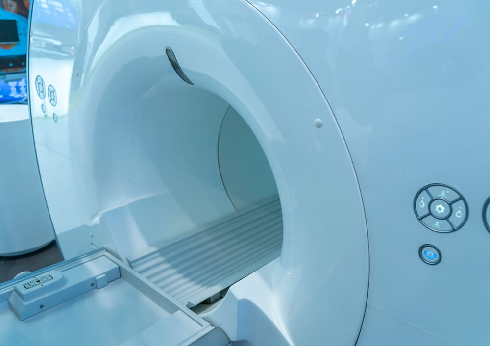
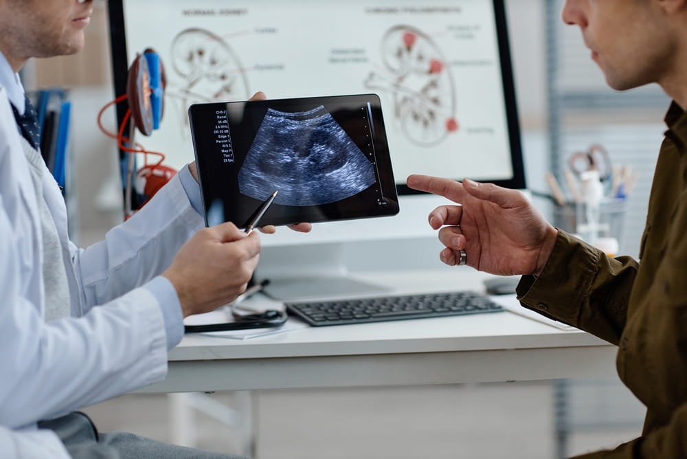
Magnetic resonance imaging provides high-resolution, cross-sectional images of soft tissue. Unlike a basic two-dimensional view, MRI can display the prostate in multiple planes and help distinguish the gland from surrounding structures. This level of anatomical detail is important when a treatment plan must account for boundaries that may be separated by only a small distance.
Before MRI-guided therapy begins, physicians can outline the prostate and identify the specific volume of tissue intended for treatment. They can also establish boundaries near the urethra, rectal wall, urinary sphincter, prostate capsule, and neurovascular structures. The resulting treatment map is based on the patient’s actual anatomy at the time of the procedure rather than an average model of prostate size and shape.
MRI may also provide information about internal prostate features. Areas affected by benign prostatic hyperplasia, previous procedures, scarring, calcification, or suspected cancer may influence how treatment is designed. While MRI findings must be interpreted alongside PSA testing, biopsy results, symptoms, and other clinical information, the images can make the treatment plan more anatomically individualized.
Real-Time Imaging Helps Confirm Positioning
A technically precise procedure begins with accurate positioning. Even a carefully designed plan may be less useful if the treatment device or the prostate is not positioned as expected. MRI guidance allows the treatment team to confirm the location of instruments and surrounding anatomy before therapeutic energy is applied.
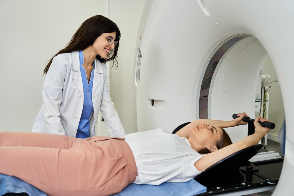
During a TULSA procedure, for example, an ultrasound applicator is placed within the urethra and positioned inside the prostate. MRI images help confirm the applicator’s relationship to the gland and the structures that need protection. The physician can then use those images to define the treatment area before ablation begins.
This confirmation step is important because the prostate is not a perfectly symmetrical organ. Its dimensions, contours, and relationship to adjacent structures differ among patients. Real-time imaging makes it possible to plan around these variations instead of assuming that the anatomy matches a standard template.
MRI Thermometry Shows Where Heat Is Traveling
One of the most significant advantages of MRI-guided thermal therapy is MRI thermometry. This technology uses changes in MRI signals to estimate tissue temperature as treatment occurs. The result is a continuously updated map showing how heat is developing and spreading within the prostate.
Temperature information matters because focused ultrasound treatment depends on delivering enough heat to ablate the planned tissue. If a region does not reach an adequate temperature, the treatment effect may be incomplete. If heat extends farther than intended, nearby healthy tissue may be exposed unnecessarily. Real-time temperature mapping gives the treatment system and physician information about both situations.
MRI thermometry differs from simply viewing the anatomy. It provides functional feedback about what the treatment energy is doing inside the tissue. The treatment team is not limited to knowing where the device is aimed; it can also observe whether the intended area is reaching the prescribed thermal range.
Tissue does not always heat uniformly. Blood flow, tissue density, gland composition, calcifications, previous treatments, and the distance between the ultrasound source and the treatment boundary can affect how energy behaves. Thermal feedback helps account for some of these variables while the procedure is still underway.
Closed-Loop Control Allows Treatment to Adapt
MRI-guided transurethral ultrasound technology can combine temperature monitoring with closed-loop control. In this type of system, the prescribed treatment boundary is established on MRI images, and software repeatedly compares measured temperatures with the planned thermal target.
The system can then modify factors such as ultrasound power, applicator rotation, frequency, or treatment speed. If tissue is heating more slowly than expected, energy delivery may be adjusted. If the treatment boundary is approaching a protected structure, the system can reduce or redirect energy based on its programming and the physician’s plan.
This adaptive process contrasts with an approach that delivers the same predetermined energy pattern regardless of how the tissue responds. Closed-loop control does not remove the physician from the procedure. Instead, it provides another layer of feedback and automation intended to keep treatment aligned with the prescribed boundaries.
The ability to respond during treatment is especially relevant because tissue changes as it heats. Swelling, altered blood flow, and coagulation may influence subsequent energy delivery. Real-time monitoring makes it possible to recognize these changes rather than discovering the final treatment pattern only after the procedure is complete.
Image Guidance Supports Protection of Nearby Structures
The prostate’s location makes precision particularly important. The urethra passes through the gland, the rectum lies directly behind it, and the urinary sphincter sits near its lower boundary. Nerves associated with erectile function run along the outer portions of the prostate. Heat extending beyond the intended area may affect one or more of these structures.
MRI guidance allows physicians to identify treatment margins in relation to these sensitive areas. The planned ablation can be shaped around the gland rather than applied as a uniform geometric volume. Protective margins may be established where preserving tissue is clinically appropriate.
Cooling technology may provide additional protection during MRI-guided transurethral ultrasound therapy. Fluid circulating through the urethral applicator helps cool the urethra, while a separate rectal cooling device may help protect the rectal wall. MRI thermometry supplements these measures by showing temperature patterns near the protected structures.
No imaging or cooling method eliminates the possibility of side effects. Urinary retention, infection, urethral narrowing, erectile changes, incontinence, rectal injury, and the need for additional treatment remain possible. Image guidance is intended to improve treatment control, not make thermal ablation risk-free.
MRI Guidance Does Not Replace Proper Diagnosis
Detailed imaging is only one part of responsible prostate treatment planning. MRI can identify suspicious areas and clarify anatomy, but it cannot independently determine every characteristic of prostate cancer or benign prostatic hyperplasia. Biopsy findings, PSA trends, prostate volume, urinary symptoms, prior treatment, overall health, and personal priorities remain essential.
A visible lesion on MRI may not represent the entire extent of clinically significant cancer. Conversely, not every MRI abnormality is cancerous. For this reason, MRI findings are interpreted alongside pathology and other clinical evidence when determining whether focal, partial-gland, or more extensive treatment is appropriate.
Patients should also understand that focused ultrasound techniques differ in how imaging is used. Some HIFU treatments use ultrasound imaging and may incorporate previously obtained MRI data or image-fusion technology. MRI-guided therapy performed inside the MRI scanner adds continuous anatomical imaging and temperature monitoring, which is different from using an MRI scan only during preprocedural planning.
More Information Does Not Guarantee a Perfect Outcome
MRI-guided treatment offers meaningful technical advantages, but the quality of a procedure still depends on patient selection, treatment planning, equipment capabilities, physician experience, and appropriate follow-up. A precise thermal map cannot compensate for an inaccurate diagnosis or a treatment plan that does not address the clinically significant disease.
Furthermore, prostate cancer may be multifocal, meaning more than one area of the gland contains cancer. A treatment focused on a visible lesion could leave separate disease untreated if diagnostic testing does not identify it. Patients considering an ablative approach should understand what portion of the prostate will be treated and why that treatment volume was selected.
Follow-up remains necessary even when MRI confirms that the intended area received thermal treatment. PSA testing, symptom monitoring, follow-up MRI, and repeat biopsy may be recommended depending on the diagnosis and treatment strategy. The prostate remains in the body after ablation and may continue producing PSA, so a single post-treatment result cannot provide a complete picture.
A More Informed Approach to Prostate Therapy
The principal advantage of MRI-guided therapy is not simply that an MRI machine is present. Its value comes from combining detailed anatomical planning, device-position confirmation, real-time temperature measurement, and the ability to adapt energy delivery according to tissue response.
Less-visible approaches may rely more heavily on fixed treatment patterns, indirect imaging, or assumptions about how energy will behave. MRI guidance gives the physician and treatment system additional information before and during therapy. That information can help define the target, evaluate thermal coverage, and establish boundaries around sensitive structures.
Patients evaluating image-guided prostate treatment should ask how the target is identified, how treatment progress is monitored, whether temperature can be measured during ablation, and how results will be evaluated afterward. Understanding these details can make discussions about the patient experience and expected follow-up more meaningful. For individuals in Farmers Branch and throughout the Dallas-Fort Worth Metroplex, Texas Prostate provides a relevant educational setting for learning how MRI guidance is changing the precision and monitoring of focused ultrasound prostate therapy.
Resources
Ghai, S., Perlis, N., Lindner, U., et al. (2023). Magnetic Resonance Imaging-Guided Ultrasound Ablation for Prostate Cancer: Contemporary Review of Performance and Outcomes. Current Opinion in Urology.
Klotz, L., Pavlovich, C. P., Chin, J., et al. (2021). Magnetic Resonance Imaging-Guided Transurethral Ultrasound Ablation of Prostate Cancer. The Journal of Urology.
Food and Drug Administration. (2019). TULSA-PRO System: 510(k) Premarket Notification K191200. U.S. Food and Drug Administration.