Advances in prostate treatment increasingly focus on addressing clinically significant tissue while reducing unnecessary disruption to the surrounding anatomy. The TULSA Procedure, short for Transurethral Ultrasound Ablation, reflects this shift by combining thermal ultrasound with detailed magnetic resonance imaging and real-time temperature monitoring. At Texas Prostate in Farmers Branch, TX, within the Dallas-Fort Worth Metroplex, understanding these features can help patients evaluate why TULSA Pro® is frequently described as a minimally invasive development in prostate tissue ablation.
The word “breakthrough” should not be interpreted as a guarantee of better results for every patient. Rather, it refers to the way several sophisticated technologies are integrated into one treatment platform. TULSA Pro does not rely on a surgical incision to reach the prostate. Instead, it delivers directional ultrasound energy from an applicator positioned inside the urethra while MRI guides and monitors the ablation.
An Inside-Out Approach to Prostate Ablation
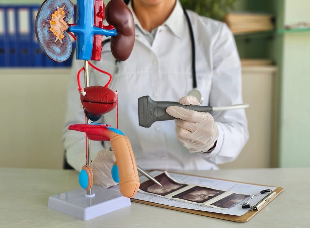
The prostate surrounds a portion of the urethra, the channel through which urine leaves the bladder. TULSA Pro takes advantage of this natural anatomical pathway. A narrow ultrasound applicator is inserted through the urethra and positioned within the prostate, allowing thermal energy to be directed outward toward selected tissue.
This approach is sometimes described as inside-out ablation. Rather than transmitting ultrasound energy through several layers of tissue before it reaches the prostate, the applicator begins near the center of the gland. Multiple ultrasound elements along the applicator can emit directional energy into different portions of the prostate as the device rotates.
The absence of a conventional surgical incision is a major reason the procedure is classified as minimally invasive. There is no need to cut through the abdominal wall or create a direct surgical opening into the prostate. However, incision-free does not mean noninvasive or risk-free. The treatment still involves anesthesia, catheter placement, thermal tissue destruction, and a period of urinary recovery.
Real-Time MRI Creates a Detailed Treatment Map
TULSA Pro is performed inside an MRI environment. Magnetic resonance imaging provides detailed views of the prostate, treatment applicator, urethra, rectum, bladder neck, urinary sphincter, and other nearby anatomy. Before energy delivery begins, the physician uses these images to outline the prostate and define the tissue intended for ablation.
This planning step is essential because treatment does not need to follow a single standard shape. Depending on the clinical objective, a physician-defined plan may target a focal area, a larger section of the gland, or nearly the entire prostate. The ability to customize the ablation boundary allows the plan to reflect the location and distribution of the tissue being addressed.
MRI also helps verify that the applicator and protective devices are properly positioned. This is important because even a carefully designed plan depends on accurate alignment between the patient’s anatomy and the equipment. The FDA-cleared system is indicated for transurethral ultrasound ablation of prostate tissue and combines MRI with directional therapeutic ultrasound and computerized process control.
Ultrasound Converts Sound Energy Into Controlled Heat
TULSA Pro does not remove prostate tissue mechanically. It uses high-energy ultrasound to heat physician-selected tissue until the cells sustain irreversible thermal damage. This process is known as thermal coagulation.
As the prostate absorbs ultrasound energy, molecular movement increases and tissue temperature rises. Once the appropriate combination of temperature and exposure time is reached, proteins lose their normal structure, cell membranes become unstable, and the targeted cells can no longer survive. The body gradually clears and remodels the damaged tissue after treatment.
The ultrasound applicator rotates during energy delivery, sweeping directional energy through the prescribed region. The system can adjust power and movement as treatment progresses rather than applying the same amount of energy throughout the entire prostate. This helps create an ablation shape that follows the physician’s treatment boundary.
Temperature Is Monitored While Treatment Occurs
One of the technology’s most distinctive features is MRI thermometry. MRI thermometry estimates temperature changes inside the prostate and surrounding tissues while ultrasound energy is being delivered. The physician and treatment software can therefore observe how the tissue is responding instead of relying exclusively on a predetermined energy setting.
This matters because prostate tissue does not heat identically in every patient. Blood flow can carry heat away from certain areas, while tissue density, fibrosis, calcifications, and prostate anatomy can affect the transmission and absorption of ultrasound. A fixed setting might produce different temperature patterns in two otherwise similar treatment plans.
TULSA Pro uses closed-loop process control to interpret temperature information and modify treatment parameters. The software can adjust ultrasound power, frequency, and applicator rotation to bring the selected tissue to the prescribed temperature. FDA documentation describes the system as automatically and dynamically regulating these factors based on real-time MR thermometry measurements.
Cooling Protects Structures Near the Treatment Area
Producing heat within the prostate is only one part of controlled ablation. The system must also limit heat exposure to structures that are not intended for treatment. This is especially important because the urethra passes through the prostate and the rectal wall lies directly behind it.
Fluid circulates through the transurethral ultrasound applicator to cool the urethra. A separate endorectal cooling device is positioned in the rectum and circulates fluid to help protect the rectal wall. Neither cooling process eliminates all risk, but both are designed to create a temperature gradient between the heated prostate tissue and adjacent protected anatomy.
The physician can also define safety boundaries around structures such as the urinary sphincter and rectum. Real-time temperature monitoring helps show whether heat is approaching those boundaries. The treatment system can then reduce or redirect energy as necessary to remain aligned with the plan.
Customization Sets It Apart From a Fixed Procedure
Prostates vary considerably in size, shape, disease location, and relationship to nearby structures. A major feature of TULSA Pro is that the treatment plan can be adapted to the individual anatomy rather than being limited to a standard ablation pattern.
For localized prostate cancer, the defined region may be based on MRI findings, biopsy results, cancer grade, disease distribution, and the location of clinically significant lesions. For BPH-related tissue ablation, planning may consider prostate volume, urinary symptoms, the location of enlarged tissue, and bladder outlet anatomy.
Customization does not mean the technology can identify abnormal cells independently. The system treats the volume selected by the physician. Diagnostic accuracy and patient selection therefore remain essential. A precise ablation device cannot compensate for disease that has not been adequately mapped or for a condition that extends beyond the intended treatment area.
Minimally Invasive Does Not Mean Minimal Follow-Up
After heat-based ablation, the prostate typically develops temporary inflammation and swelling. Because the gland surrounds the urethra, this response can interfere with urinary flow during the early recovery period. A catheter is generally used to allow urine to drain while swelling begins to improve.
The ablated tissue does not disappear immediately. Damaged cells are gradually cleared, and the treated area changes as healing and tissue remodeling occur. Temporary urinary frequency, urgency, irritation, blood in the urine, pelvic discomfort, or changes in urinary flow may occur during recovery.
The broader patient experience can include preparation, anesthesia, treatment inside the MRI suite, catheter management, recovery instructions, and subsequent monitoring. Depending on the original diagnosis, follow-up may involve symptom assessments, PSA testing, imaging, urinary testing, or biopsy.
Why Patient Selection Still Matters
Not every patient with prostate cancer or BPH is automatically an appropriate candidate for TULSA Pro. Prostate calcifications may interfere with the passage of ultrasound energy. Gland dimensions, urethral anatomy, previous procedures, MRI compatibility, cancer location, disease risk, and medical history can also affect eligibility.
The possible benefits must be considered alongside treatment limitations and uncertainties. Research has demonstrated the feasibility of MRI-guided transurethral ultrasound ablation and its ability to produce physician-defined treatment patterns, but long-term outcomes and comparative evidence continue to develop. Studies of localized prostate cancer have therefore emphasized careful patient selection and continued surveillance after treatment.
A thorough evaluation should clarify what portion of the prostate would be treated, why that volume has been selected, which structures would be protected, and how treatment response would be measured. Patients should also understand the possibility of persistent disease, recurrent symptoms, side effects, or the need for additional care.
A Coordinated Technological Advance
What makes TULSA Pro a minimally invasive breakthrough is not one isolated feature. Its distinction comes from the coordination of several components: a natural transurethral access route, directional thermal ultrasound, detailed MRI planning, live temperature mapping, automated energy adjustments, and protective cooling.
Together, these components allow the physician to plan and monitor a customized prostate ablation without making a conventional surgical incision. The technology does not remove the complexities of prostate disease, and it does not replace careful diagnosis, informed decision-making, or long-term follow-up. It does, however, provide a highly controlled method for delivering thermal energy into a selected region of the prostate.
For individuals researching minimally invasive prostate tissue ablation in the Dallas-Fort Worth Metroplex, Texas Prostate in Farmers Branch, TX, provides a local point of context for learning how TULSA Pro combines MRI, directional ultrasound, thermal feedback, and individualized planning.
Resources
Chin, J. L., Billia, M., Relle, J., Roethke, M. C., Popeneciu, I. V., Kuru, T. H., Hatiboglu, G., Mueller-Wolf, M. B., Motsch, J., Schlemmer, H. P., & others. (2016). Magnetic Resonance Imaging-Guided Transurethral Ultrasound Ablation of Prostate Tissue in Patients With Localized Prostate Cancer: A Prospective Phase 1 Clinical Trial. European Urology.
Klotz, L., Pavlovich, C. P., Chin, J., Hatiboglu, G., Koch, M., Penson, D., Kaye, D., Crouzet, S., Villers, A., & others. (2021). Magnetic Resonance Imaging-Guided Transurethral Ultrasound Ablation of Prostate Cancer. Journal of Urology.
U.S. Food and Drug Administration. (2019). TULSA-PRO System: 510(k) Premarket Notification K191200. U.S. Department of Health and Human Services.