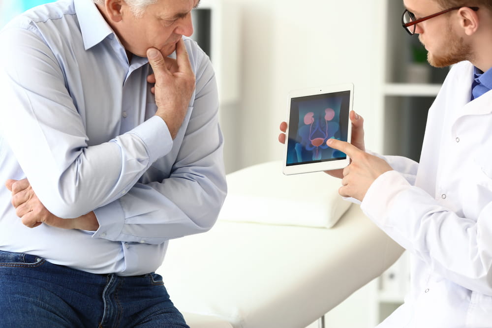
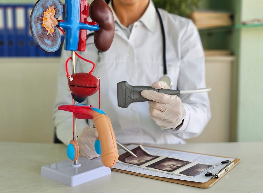
Heat-based ablation uses carefully controlled thermal energy to damage a selected volume of prostate tissue. Instead of removing tissue directly, ablation raises its temperature until the cells can no longer survive. Imaging, treatment-planning software, energy-delivery devices, and protective cooling may all work together to shape the treatment area. At Texas Prostate in Farmers Branch, TX, within the Dallas-Fort Worth Metroplex, understanding this science can help patients make sense of focused ultrasound approaches considered for localized prostate cancer and benign prostatic hyperplasia, or BPH.
The idea of using heat to alter biological tissue is not new. What has changed is the degree of control available through modern imaging and therapeutic systems. Physicians can define a treatment boundary, deliver energy into that region, monitor how the tissue responds, and take steps to reduce heat exposure to nearby structures. The result is not simply “heating the prostate,” but creating a planned thermal dose within a specific anatomical area.
How Heat Causes Irreversible Cellular Damage
Human cells function within a relatively narrow temperature range. When tissue temperature rises above normal levels, proteins begin to change shape and lose their ability to perform essential cellular functions. Cell membranes become unstable, enzymes stop working normally, and the structures responsible for maintaining and repairing the cell begin to fail.
The effect depends on both temperature and time. Moderate heating sustained for a longer period can damage tissue, while a higher temperature may produce an irreversible effect within seconds. Many thermal ablation techniques are designed to raise targeted tissue to temperatures that cause coagulative necrosis, a form of cell death associated with protein denaturation and structural damage. Research on focused ultrasound commonly describes temperatures in the approximate range of 60 to 90 degrees Celsius within the focal region, although the actual treatment parameters depend on the system and plan being used.
The objective is to deliver enough thermal energy to create a predictable zone of nonviable tissue. If a region does not receive a sufficient combination of heat and exposure time, some cells may survive. If heat extends beyond the planned boundary, nearby tissue may be affected. This is why successful ablation depends on controlling the total thermal dose rather than merely reaching a single peak temperature.
How Focused Ultrasound Creates Heat
Ultrasound consists of mechanical sound waves that travel through tissue. Diagnostic ultrasound uses relatively low energy to create images from returning echoes. Therapeutic ultrasound uses substantially greater energy and directs it toward a treatment target.
With HIFU, or high-intensity focused ultrasound, multiple ultrasound waves converge within a small focal region in the prostate. Each individual wave passes through intervening tissue with less intensity, but their combined energy becomes much greater at the focal point. The tissue absorbs that energy, molecular motion increases, and heat develops rapidly.
The focal zone is relatively small, so treatment is performed in a sequence. The system directs energy to one location and then advances to the next planned position. The overlapping focal zones eventually form a larger ablation volume. The U.S. Food and Drug Administration defines this type of device as an image-guided system that transmits high-intensity therapeutic ultrasound energy into the prostate to thermally ablate a defined, targeted tissue volume.
Thermal Effects Beyond Basic Heating
Heat is the primary mechanism behind focused ultrasound ablation, but intense acoustic energy can also create mechanical effects. One such effect is cavitation, in which microscopic gas bubbles form, move, or collapse within tissue fluids. These changes may place additional mechanical stress on nearby cells.
The relative contribution of cavitation can vary with ultrasound frequency, energy intensity, pulse duration, tissue properties, and the design of the treatment system. In controlled prostate ablation, the physician is generally seeking a predictable thermal effect rather than uncontrolled bubble activity. Treatment settings are therefore selected to create a stable ablation zone while limiting unpredictable tissue disruption.
Blood circulation also affects how tissue heats. Flowing blood can carry thermal energy away from a region, a process sometimes called the heat-sink effect. Tissue located near larger blood vessels may consequently warm differently from tissue with less circulation. Prostate composition, fibrosis, calcifications, and previous treatment can also influence how ultrasound energy travels or is absorbed.
Why Image Guidance Is Essential
The prostate sits close to several structures that influence urinary, sexual, and bowel function. The urethra travels through the gland, the urinary sphincter lies near its lower portion, the bladder neck is positioned above it, and the rectum is directly behind it. Neurovascular structures also run near the outer prostate.
Because these structures are close together, the physician must understand exactly where the treatment energy is being placed. Imaging helps identify the prostate’s boundaries, the intended treatment zone, the position of the energy-delivery device, and areas that require additional protection.
Ultrasound-guided systems may use real-time ultrasound imaging to align the focal point with the treatment plan. Other approaches rely on magnetic resonance imaging. The TULSA Procedure, whose name stands for Transurethral Ultrasound Ablation, uses an applicator positioned within the urethra and MRI guidance to direct ultrasound energy outward into selected prostate tissue.
How Temperature Monitoring Shapes the Treatment Zone
Knowing where energy is directed is important, but location alone does not reveal how hot the tissue has become. Temperature monitoring provides another layer of information by showing whether the target is receiving the intended thermal dose.
MRI thermometry estimates temperature changes as treatment proceeds. A computer can compare the measured temperature pattern with the physician-defined treatment boundary. If an area is not warming sufficiently, energy delivery may be adjusted. If heat approaches a protected margin, the system may reduce power, change applicator movement, or stop energy delivery in that direction.
This creates a feedback loop rather than a fixed application of heat. The treatment plan defines the target, the device delivers ultrasound, temperature data reveal the tissue response, and the system modifies delivery according to that information. Because biological tissue does not heat identically in every patient, real-time feedback can improve control over the final ablation pattern.
The Role of Cooling and Safety Margins
A thermal ablation system must manage heat outside the target as carefully as it creates heat within the target. Cooling mechanisms may protect structures that are close to the energy source or treatment boundary.
In transurethral ultrasound ablation, fluid circulating through the applicator can help protect the urethra even though the device is positioned inside it. A separate cooling device placed in the rectum may help limit temperature increases along the rectal wall. These protective measures establish a temperature gradient in which the selected prostate tissue is heated while adjacent structures are actively cooled.
Physicians may also establish safety margins around the urinary sphincter, rectum, bladder neck, or neurovascular structures. These margins can influence how much tissue is treated and how closely the ablation boundary approaches sensitive anatomy. Precision therefore involves both active energy delivery and deliberate areas of reduced or avoided heating.
What Happens to Tissue After Ablation
Heat-damaged tissue does not immediately vanish. Directly after treatment, the affected cells lose function, local blood vessels may close, and inflammation begins. Swelling can temporarily increase pressure around the urethra, which is one reason catheterization may be needed during the early recovery period.
Over the following weeks and months, the body gradually clears damaged cells and remodels the treated region. Fibrosis and scar-like changes may develop as healing progresses. The prostate’s final size and shape may therefore evolve rather than changing completely on the treatment day.
The extent of this response depends partly on the amount and location of tissue treated. A focal plan may ablate a limited lesion and surrounding margin, while partial-gland or whole-gland plans involve larger volumes. Focused technology does not inherently mean that only one tiny area must be treated; it means energy can be directed according to a physician-defined treatment pattern.
Why Treatment Planning Remains Individualized
Heat cannot independently recognize whether a prostate cell is cancerous, enlarged, inflamed, or healthy. Instead, diagnostic information is used to determine which tissue should be included within the treatment boundary. MRI findings, biopsy results, PSA measurements, prostate size, symptom severity, disease distribution, and previous treatment history may all influence that decision.
Ablation is therefore only one part of a broader clinical process. The precision of an energy-delivery system cannot compensate for an incomplete diagnosis or an unsuitable treatment plan. Current clinical guidance also notes that patients considering focal or whole-gland ablation for localized prostate cancer should understand the limitations of long-term comparative evidence and the importance of appropriate selection and surveillance.
Treatment does not eliminate the need for follow-up. The ongoing patient experience may include urinary recovery, catheter care, symptom monitoring, PSA testing, imaging, or biopsy. Follow-up helps determine whether the treated area responded as expected and whether untreated prostate tissue requires continued observation.
Precision Depends on an Entire System
The science of heat-based prostate ablation involves more than raising tissue temperature. It requires an energy source capable of reaching the prostate, imaging that defines the anatomy, software that shapes the treatment zone, temperature information that reflects tissue response, and cooling or safety margins that help protect nearby structures.
Every part of this process contributes to the final result. The ultrasound applicator produces energy, but the treatment plan determines where it should go. Heat creates cellular damage, but monitoring helps determine whether the thermal dose is adequate. Cooling does not treat the prostate, but it supports the effort to confine treatment to the intended region.
For patients in the Dallas-Fort Worth Metroplex learning about focused ultrasound, Texas Prostate in Farmers Branch, TX, provides a local context for understanding how image guidance, thermal energy, feedback systems, and individualized planning come together in heat-based prostate tissue ablation.
Resources
Ashrafi, A. N., Nassiri, N., Gill, I. S., & Djaladat, H. (2019). Ablation Energies for Focal Treatment of Prostate Cancer. World Journal of Urology.
Ghai, S., Perlis, N., Lindner, U., Haider, M. A., & others. (2018). Magnetic Resonance Guided Focused High Frequency Ultrasound Ablation for Focal Therapy in Prostate Cancer. Journal of Urology.
U.S. Food and Drug Administration. (2015). De Novo Classification Request for the Sonablate 450 High Intensity Ultrasound System for Prostate Tissue Ablation. U.S. Department of Health and Human Services.